Neurobiology
Many studies link BPD with brain areas related to emotions, like the amygdala, insula, posterior cingulate cortex, hippocampus, anterior cingulate cortex, and prefrontal regions that regulate emotions.
People with BPD might experience an irregularity in their brain chemistry, specifically related to neurotransmitters like serotonin, dopamine, and norepinephrine. These chemical signals have a crucial impact on managing mood, controlling impulses, and handling emotional reactions.
DBT has been shown to reduce amygdala hyperactivity, which is associated with improvements in emotion regulation and increased use of emotion regulation strategies. These findings suggest that dysfunctional circuits involving hyperactive limbic regions and hypoactive prefrontal modulation, particularly in the dorsal lateral prefrontal cortex, are the anatomical correlates of BPD.


Neurotransmitters & biology
Oxytocin
Oxytocin plays a role in regulating social cognition through the frontolimbic system. People with BPD have been found to have structural and functional differences in this system. In facial recognition studies, individuals with BPD tend to perceive negative and untrustworthy emotions more often. This suggests that oxytocin's influence on the salience network contributes to interpersonal hypersensitivity in BPD.
Oxytocin also helps regulate the hypothalamic-pituitary-adrenal axis, which helps to reduce fear and extinguish the startle response to previously emotionally charged stimuli. Additionally, oxytocin's impact on attachment and affiliative systems may contribute to the anger, impulsivity, and emotional instability seen in individuals with BPD in response to perceived insults.

Related to the serotonergic system, dopamine dysfunction has been suggested as a factor in BPD symptoms as well. Dopamine is thought to play a role in emotional information processing and impulsivity, as well as general cognition.
Dopamine

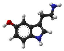
Serotonin is one of the neurotransmitters implicated in normal personality.
BPD is characterized by symptoms related to serotonin imbalance, such as mood swings, suicidal tendencies, impulsiveness, and lack of self-control. Studies show that selective serotonin re-uptake inhibitors are effective in managing depression and impulsivity in BPD patients.

Serotonin
Amygdala


Borderline personality disorder can lead to a highly sensitive automatic nervous system, making the sympathetic nervous system easily triggered. Adrenal glands release adrenaline and norepinephrine, causing a 'flight ot fight' response.
This can result in exaggerated or irrational responses, with individuals experiencing prolonged stress compared to those without BPD.
Norepinephrine

In mood and emotion disorders, the amygdala is frequently hyperactive and plays a crucial role in regulating vigilance and generating negative emotional states.
Research has indicated that individuals with BPD may have a reduction in the size of their hippocampus and amygdala by up to 16%. It has been proposed that traumatic experiences could potentially contribute to these changes in brain structure.
Hippocampus

The hippocampus is important for controlling emotions and memories, and is involved in the development of BPD.
People with BPD have smaller hippocampal size, different activation in the hippocampus, and problems connecting with other parts of the brain. These issues are connected to various symptoms of BPD, like difficulty managing emotions, acting impulsively, and having an unstable self-perception.
Neuro-Imaging
In recent years, neuroimaging has emerged as a crucial tool in clinical neurobiology, playing a pivotal role in understanding the brain. It encompasses various techniques like positron emission tomography (PET), functional and structural magnetic resonance imaging (fMRI), MR spectroscopy, and diffusion tensor imaging (DTI).


Functional MRI detects brain activity by monitoring changes in blood flow. This method works because blood flow and neuronal activity are linked. When a specific brain area is active, blood flow to that area rises.
fMRI
PET
MR Spectroscopy
DTI
A PET scan is an imaging test that uses a radioactive tracer to detect disease in the body and show how organs and tissues are functioning.
MR spectroscopy is a diagnostic test that can measure biochemical changes in the brain without being invasive. It is particularly useful for detecting tumors by comparing the chemical composition of normal brain tissue to that of abnormal tumor tissue.
Diffusion tensor imaging tractography, or DTI tractography, is an MRI (magnetic resonance imaging) technique that measures the rate of water diffusion between cells to understand and create a map of the body's internal structures.


